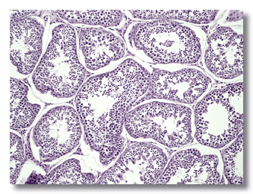
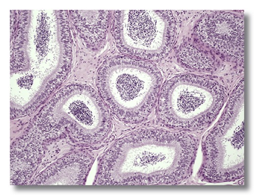
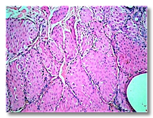
1. TESTIS
It is surrounded by the tunica albuginea, a solid capsule of dense irregular connective tissue, continuous with connective tissue trabeculae, the septula testis.
The convoluted seminiferous tubules are lined by stratified epithelium consisting of spermatogenic cells and sustentacular Sertolli cells. The interstitial endocrine cells (Leydig cells) are irregular, polyhedral cells and contain lipid inclusions.
The ductuli efferentes are lined by a simple columnar epithelium with ciliated and nonciliated cells.
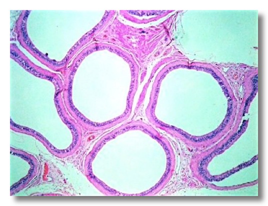
The ductus epididymidis is extremely tortuous and is lined by a pseudostratified epithelium with stereocilia.
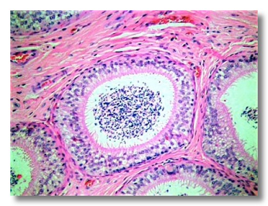
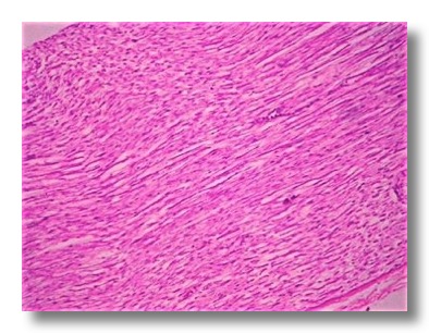
The ductus deferens has a well developed tunica muscularis.

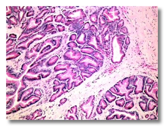
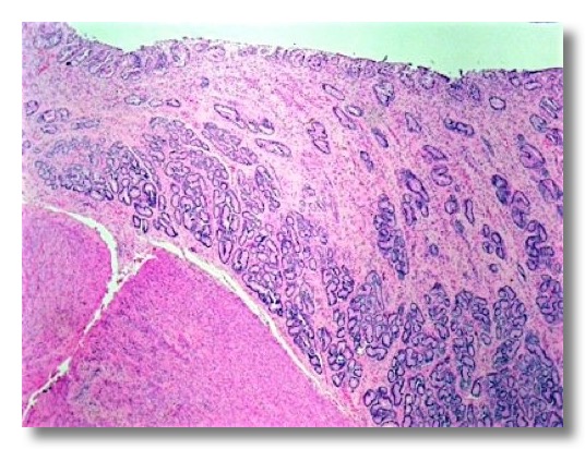
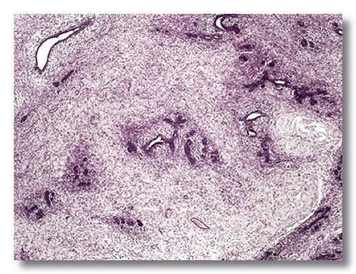
2. PROSTATE
The prostate consists of a varying number of individual tubuloalveolar glands lined by a simple or pseudostratified columnar epithelium (depending on the species). Secretory portions and ducts of the prostate are surrounded by loose connective tissue containing smooth muscle cells. In the dog the secretion is serous, whereas in other animals it is seromucous.
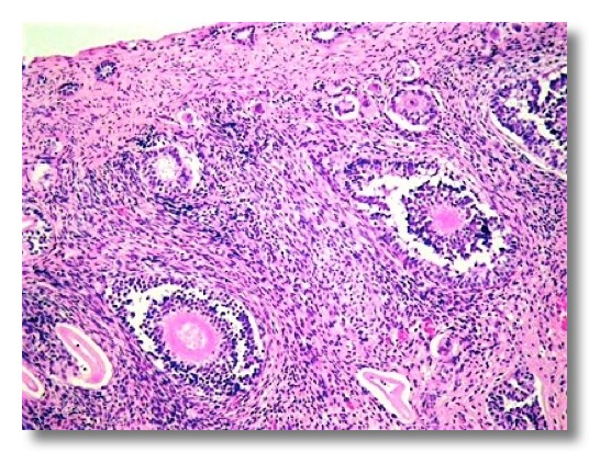
3. OVARY
It is divided into an outer cortex and an inner medulla. The cortex is covered by a low cuboidal surface epithelium. The tunica albuginea is a thick connective tissue layer immediately beneath the surface epithelium. The cortex contains follicles and corpora lutea. In the cortex we can observe: primordial follicles (composed of an oocyte surrounded by a single layer of squamous epithelial cells), primary follicles (oocyte, simple cuboidal epithelium), secondary follicles (oocyte, zona pellucida, stratified epithelium of polyhedral granulosa cells), vesicular secondary follicles (with small fluid-filled cavities among the granulosa cells), tertiary or Graafian follicles (oocyte surrounded by the corona radiate, granulosa cells, antrum, theca interna and theca externa), and corpus luteum (formed by luteal cells derived from the granulosa and theca cells).
 .
. 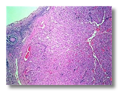
In the avian ovary, large follicles are suspended from the surface of the ovary by stalks of cortical tissue. Each follicle consists of a growing, yolk-laden oocyte with a rounded nucleus.
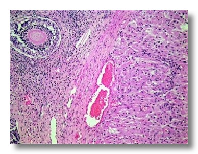
4. UTERUS
The uterine mucosa is called endometrium. Its epithelial lining is simple cuboidal or columnar in the mare and carnivores, and pseudostratified in the sow and ruminants. The lamina propria-submucosa contains numerous simple tubular endometrial glands that open into the lumen of the uterus.
The myometrium (muscularis) is composed of a deep inner layer of circular smooth muscle and a less clearly defined outer layer of longitudinal smooth muscle. A stratum vascular is also observed.
The perimetrium (tunica serosa) consists of loose connective tissue covered by the peritoneal mesothelium.
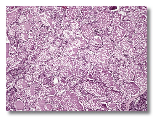
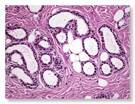
5. MAMMARY GLAND
The mammary gland is a compound tubuloalveolar gland. Groups of tubuloalveolar secretory units form lobules separated by connective tissue septa. The secretory unit is lined by a simple cuboidal epithelium that varies in height during various stages of secretory phase. Myoepithelial cells lie between the secretory cells and the basement membrane.
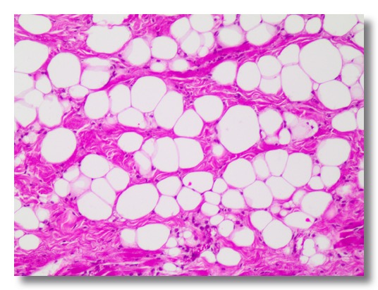
In the non-lactating gland, the lobules consist of a duct system surrounded by loose connective tissue, separated from the adjoining lobules by fatty interlobular tissue. In the lactating gland, the duct system expands and the alveoli are filled with milk secretion.
The duct system is lined by a simple cuboidal or columnar epithelium without secretory activity.
 .
. 
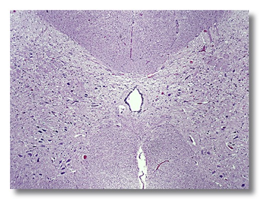
6. SPINAL CORD
Spinal cord consists of gray matter and white matter, as the other organs of the central nervous system. The gray matter is H-shaped and has bilateral small dorsal columns (horns) and large ventral columns. Gray matter is rich in neuronal cell bodies, and glial cells. The central canal is lined by a simple cuboidal epithelium (ependyma).
White matter is formed by dense accumulations of axons. Glial cells are also observed.
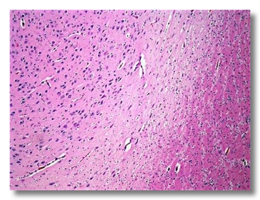
7. CEREBRUM
The pia mater meninge coats the entire cerebrum surface. Gray matter on the surface of cerebrum is called cortex. Gray matter accumulations within the cerebral white matter are called nuclei. In mammals, the cerebral cortex is divisible into six layers. Neuronal cell bodies and glial cells are detected.
The white matter is homogeneous, without neuronal cell bodies, and contains myelinated and nonmyelinated axons and oligodendrocytes.
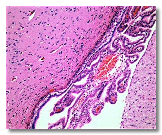
Cerebrospinal fluid is produced by choroid plexuses located in the brain ventricles. The plexus consists of dense microvasculature in loose connective tissue coated by modified ependymal cells, called choroid plexus epithelium.
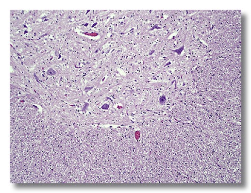
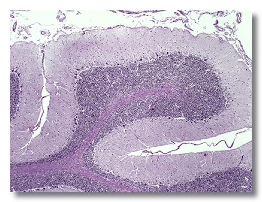
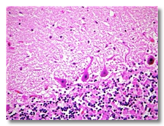
8. CEREBELLUM
The cerebellar cortex (gray matter) can be divided into three layers. The molecular layer, composed predominantly of neuropil, is most superficial. The Purkinje cells send axons into the white matter and dendritic trees that project into the molecular layer. The granule cell layer, situated adjacent to white matter, features densely packed granule cells.
White matter is deep to the cortex, and cerebellar nuclei are embedded within the white matter.
 .
. 
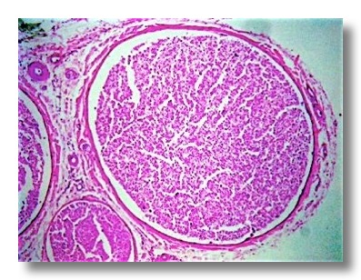
9. PERIPHERAL NERVOUS SYSTEM
Cyatic nerve, chicken
Nerves are composed of one to several nerve fascicles. A nerve fascicle is delimited by perineurium. The multiple fascicles of a nerve are bound together by epineurium, a connective tissue less dense than fibrous perineurium. The nerve fascicles contain thousands of myelinated and nonmyelinated axons. Schwann’s cells are also detected.
 .
. 
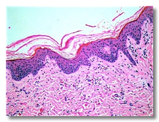
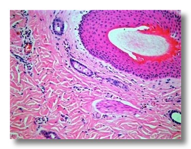
10. SKIN
The epidermis is a keratinized stratified squamous epithelium. At least four layers can be identified: stratum basale, stratum spinosum, stratum granulosum, and stratum corneum. The thickness of the epidermis and degree of keratinization vary with its anatomical location.
The dermis is generally divided into a superficial (papillary) (loose connective tissue) layer and a deep (reticular) (dense irregular connective tissue) layer. The papillary layer forms projections called dermal papillae. Dermal papillae are reduced or absent in thin skin.
Hair follicles are seen at various levels throughout the dermis. Bundles of smooth muscle cells form the arrector muscle (arrector pili). Sebaceous glands are associated with the hair follicles; they are holocrine glands formed by a solid mass of epidermal cells.
 .
. 
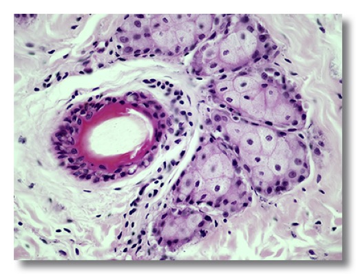
Apocrine glands are simple saccular or tubular glands with a large lumen lined by a cuboidal epithelium. Merocrine glands are found mainly in the footpads of carnivores.
The hypodermis (subcutis) is a layer of connective tissue that anchors the dermis to the underlying muscle. Adipose tissue is present forming the panniculus adiposus.
11. PERIANAL (CIRCUMANAL) GLANDS
These glands are lobulated, modified sebaceous glands located in the dermis around the anus of dogs. They are composed of solid, compact masses of polygonal cells resembling closely packed liver cells (hepatoid glands).
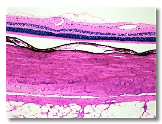
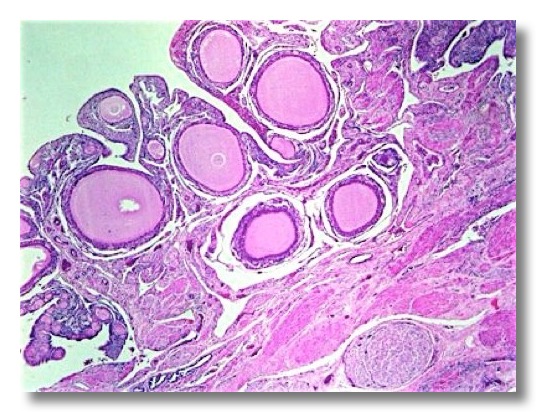
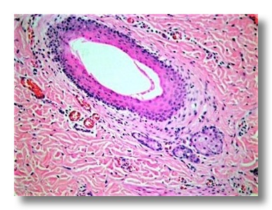
12. EYE
The cornea is lined by a nonkeratinized stratified squamous epithelium resting on a basement membrane (Bowmann’s). The underlying stroma (substantia propria) consists of dense connective tissue. The caudal limiting membrane (Descemet’s) separates the substantia propria from the posterior epithelium (simple squamous corneal endothelium).
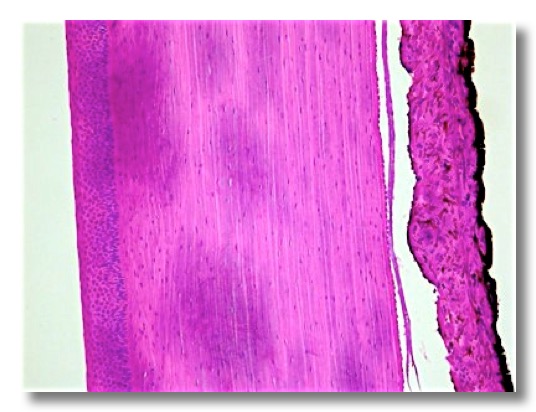
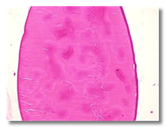
The lens is a biconvex body composed of epithelial cells within a homogeneous outer capsule. The anterior epithelial cells are cuboidal at the pole, and become elongated, prismatic and arranged in meridional rows at the equator where they form lens fibers.
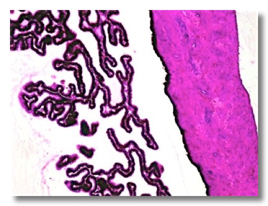
The ciliary processes are lined by a basal layer (pigmented columnar cells) and a surface layer (nonpigmented columnar cells). The iris consists of a stroma of pigmented, highly vascularised loose connective tissue, the sphincter and dilator muscles, and the posterior (pigmented) epithelium. The anterior surface is non-epithelial, and is covered by fibrocytes and melanocytes.
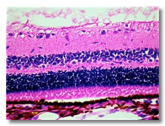
The retina is the innermost layer of the wall of the eye. From the choroid to the cavity of vitreous humour 10 layers are identified.