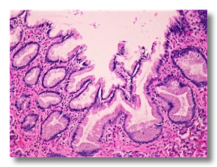
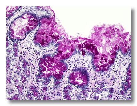
1. STOMACH
The tunica mucosa is composed of simple columnar mucous secretory epithelium, a lamina propria with gastric glands, and a muscularis mucosae. The gastric glands occupy most of the lamina propria, so that only a few connective tissue fibers and cells are seen between them. Four cell types are present in the fundic glands: mucous neck cells, chief cells, parietal cells, and endocrine cells.
The chief cells or zymogen cells are basophilic and secrete pepsinogen. The parietal cells are eosinophilic and secrete ClH.
The submucosa contains collagen fibers, fat, and the submucosal nerve plexuses.
The tunica muscularis has three layers: an inner oblique, a middle circular, and an outer longitudinal layer. The myenteric plexuses are located between the middle and outer muscle layers.
The tunica serosa is composed of mesothelium overlying a layer of loose connective tissue.
 .
. 
 .
. 
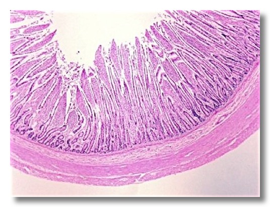
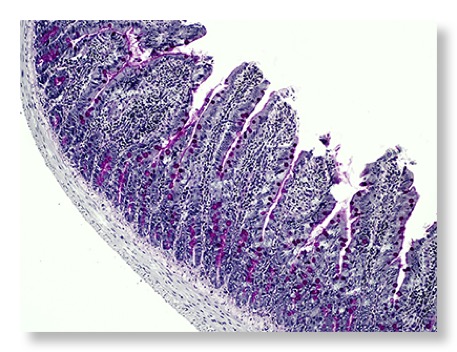
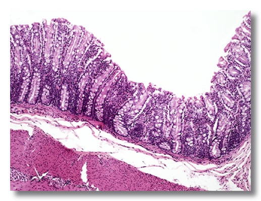
2. SMALL INTESTINE
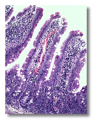
The villi are mucosal projections and are the most characteristic feature of the small intestine. The intestinal crypts, which open between the bases of the villi, penetrate the mucosa as far as the muscularis mucosae. The lumen of the intestine is lined by simple columnar epithelium with numerous goblet cells interspersed among the columnar cells. The columnar absorbing cells have oval nuclei situated near the cell base and have prominent microvilli (striated border). The lamina propria and muscularis mucosae can be easily identified.
The submucosa is composed of loose connective tissue. Submucosal glands of variable nature (mucous, serous or seromucous) can be observed although their distribution varies with the different species. In the ileum Peyer’s patches can be observed.
The tunica muscularis consists of inner circular and outer longitudinal smooth muscle layers.
The tunica serosa is composed of mesothelium overlying a layer of loose connective tissue.
 .
. 
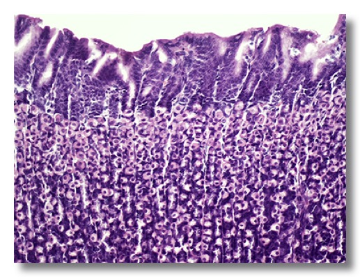
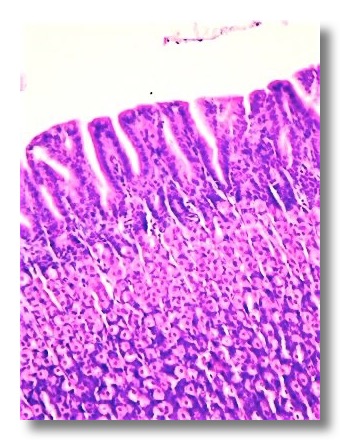
3. LARGE INTESTINE
The epithelium is also simple columnar epithelium but it is remarkable the high number of goblet cells. It is also characteristic the absence of villi, common to all segments of the large intestine. There are longer, more compact intestinal crypts, with large numbers of goblet cells.
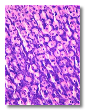
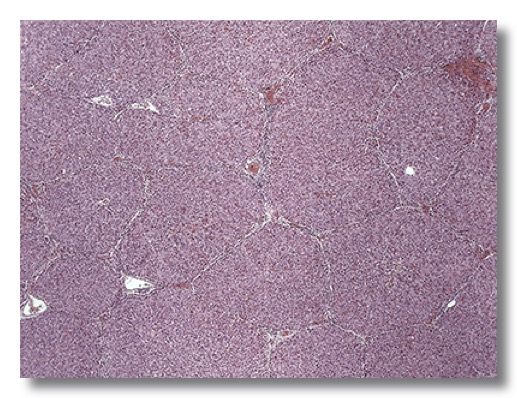
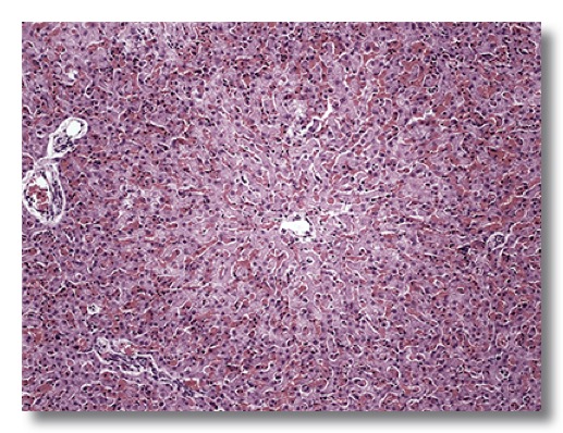
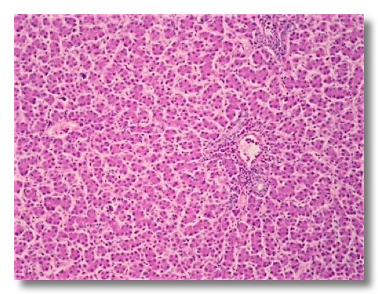
4. LIVER (PIG)
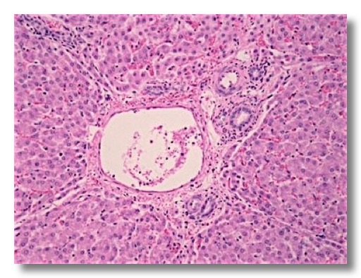
Liver is covered by visceral peritoneal mesothelial cells overlying a thin connective tissue capsule. Connective tissue from the capsule extends into the interlobular spaces, difficult to see, except in the pig, which has distinct fibrous connective tissue septa surrounding each liver lobule. The hepatic lobule is organized around the central vein, and cross-sectional profiles are hexagon-shaped, with the sinusoids radiating from the periphery to the central vein into which they empty. Portal areas are present at some angles of the hepatic lobules. Portal areas contain branches of the hepatic artery (arterioles), portal vein (venules) and the bile interlobular ductule.
The hepatocytes are polyhedron-shaped cells with a centrally located spherical nucleus (occasionally are seen binucleated hepatocytes). Sinusoids are lined by a discontinuous endothelium and macrophages (Kupffer cells).
 .
.
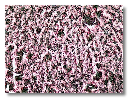
A network of fine reticular fibers provides a supportive framework.
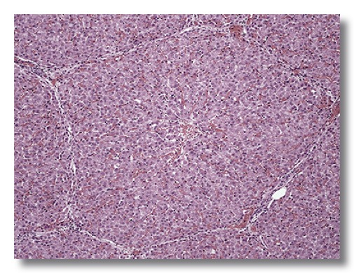
5. LIVER (OTHER SPECIES)
Liver, dog
The histological structure is similar, but interlobular septa (and thus the hepatic lobules) are difficult to see.
Liver, avian species
The hepatocytes are arranged in rows, often two cells thick, separated by sinusoids.
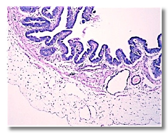
6. GALLBLADDER
When empty the gallbladder mucosa is thrown into numerous folds; as it fills and expands, the folds tend disappear. The gallbladder is lined by a simple columnar epithelium. Goblet cells are characteristic of the epithelium of some species (cattle).
Because there is no muscularis mucosae, the lamina propria blends with the submucosa without a clear line of junction.
The tunica muscularis is thin and composed of circular smooth muscle fibers.
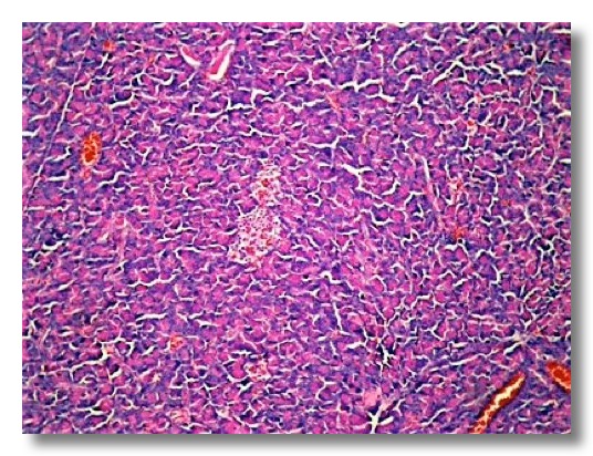
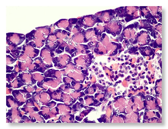
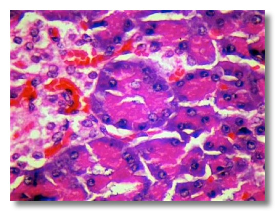
7. PANCREAS
The stroma of the pancreas consists of a thin capsule that gives rise to delicate connective tissue septa separating the parenchyma into distinct lobules.
Exocrine pancreas
The secretory units are tubuloacinar serous units. The secretory epithelial cells are pyramid-shaped, with a spherical nucleus near the base of the cells. The cytoplasm surrounding the nuclear region is basophilic, whereas the apical region of the cells contains eosinophilic zymogen granules.
Each secretory unit has a small lumen, and the duct system begins as flattened centroacinar cells in the lumen. These cells begin in the acinar portion and continue throughout the tubular part of the secretory unit. The ductal epithelium changes from cuboidal in the interlobular ducts to columnar in the interlobular and collecting ducts.
Endocrine pancreas
The endocrine cells of the pancreas are clustered in pancreatic islets (Langerhans’ islets). The islet cells are arranged in irregular anastomosing cords composed of different cell types. The pancreatic islets are supplied by a dense capillary network.